Roof Of Pterygomandibular Space Is Formed By

The Roof Of Pterygomandibular Space Is Formed By

The Roof Of Pterygomandibular Space Is Formed By

Physician Assistant Pa 2013 Session 1 Tsai Flashcards An3 07 Nasal Cavity Parasinuses And Nasopharynx Studyblue Nasal Cavity Cavities Oral Cavity

Test Fasciae And Spaces H N Ii Quizlet
Anatomy Of The Pterygomandibular Space And Its Clinical Significance

Oral Cavity And Mastication Flashcards Quizlet

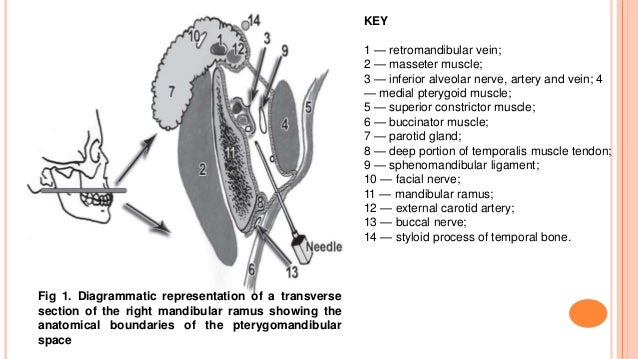
Posteriorly parotid glandular tissue curves medially around the back of the mandibular ramus to form a posterior border while anteriorly the buccinator and superior constrictor muscles come together to form a fibrous junction the pterygomandibular raphe.
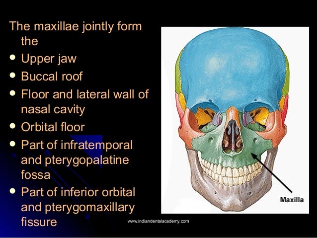
Roof of pterygomandibular space is formed by.

Pterygopalatine Fossa Anatomy Arterial Supply Venous Drainage Nerve Supply Radiology Dental Science In 2020 Anatomy Fossa Maxillary Sinus

Fascial Space Infections

Pharynx Flashcards Quizlet
Anatomy Of The Pterygomandibular Space And Its Clinical Significance

Oral Cavity Flashcards Quizlet

Sensitive Teeth Are Primarily Caused By Gum Recession Gums Recede For Many Reasons Like Brushing Too Hard Age And Pe Dental Dental Hygiene School Dentistry
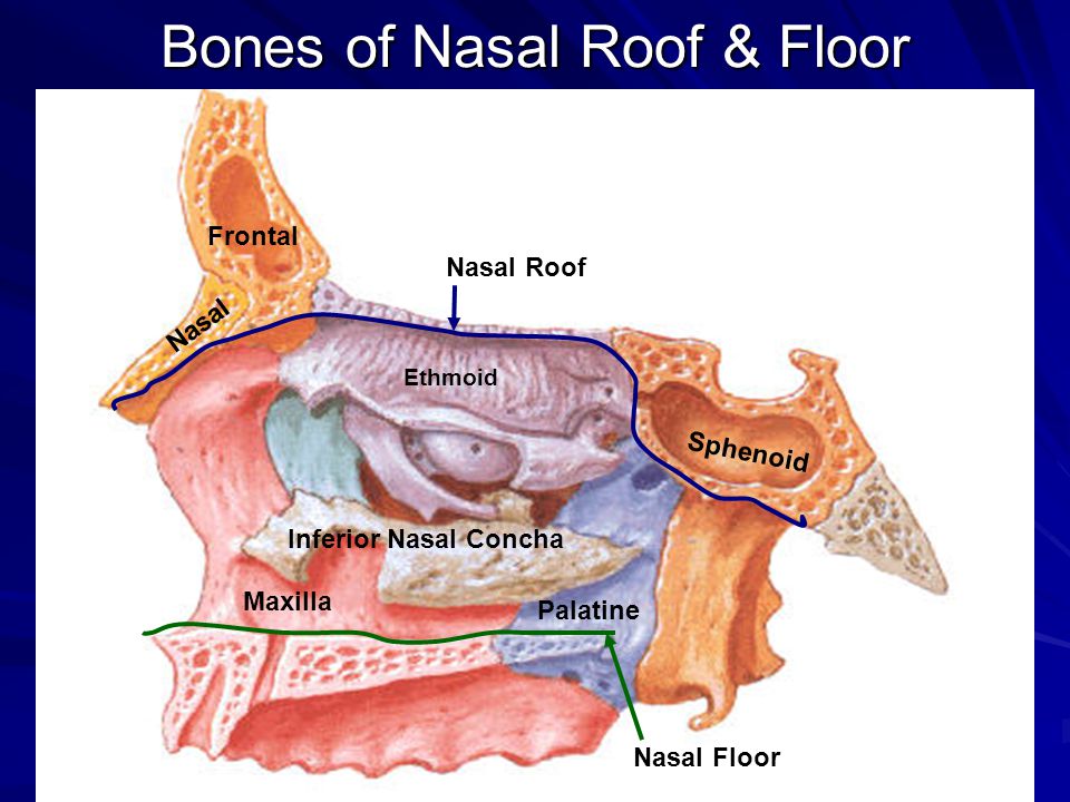
Gross Anatomy Of The Nasal Cavity The Pharynx Ppt Video Online Download
Biological Considerations Of Maxillary And Mandibular Impressions

Forces Of Occlusion

The Underused Block The Vazirani Akinosi Mandibular Block Is A Viable Pain Control Option Registered Dental Hygienist Rdh Magazine

Imagem Relacionada Goruntuler Ile

Clinical Implications Of Growth And Development Certified Fixed O
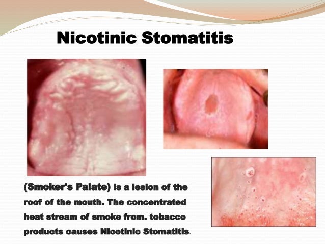
Tongue Softpalate Floor Of The Mouth Cosmetic Dentistry Courses

Pin Auf Travel
10 Post Insertion Problems And Complaints

A Glimpse Of Past The Temporo Buccinator Band Of Hovelaque Or The Buccotemporal Fascia Of Zenker Sciencedirect

The Digestive System In The Head And Neck Ppt Video Online Download

Muscles Osteology Anatomy Physiology Pharmacology 200 With Me At Tufts University Studyblue
1
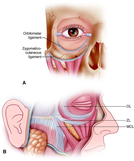
Anatomy Of The Midface Springerlink

Anatomy Respect In Implant Dentistry Assortment Location Clinical Importance Review Article

Salivary Gland Anatomy Pocket Dentistry
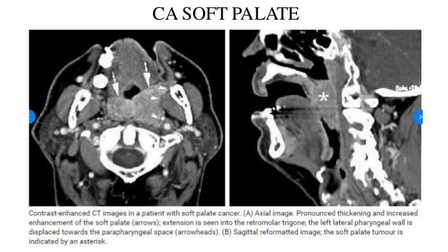
Radio Logical Anatomy Of Head And Neck Cancers
Http Link Springer Com Content Pdf 10 1007 2f978 1 4614 4466 4 Pdf
Source : pinterest.com
